Vole Photo Gallery |
|
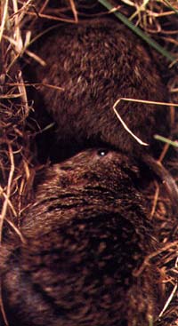
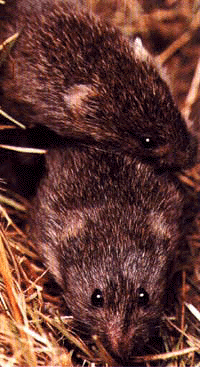
A male and female prairie vole |
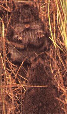
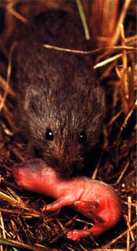
A male prairie vole retrieves a pup |
![]() |
![]() |
Exposure to an unrelated male or its urinary pheromone is essential to induce estrus in female voles. |
Male prairie voles become aggressive after mating and exhibit aggression towards intruders. |
A virgin female sniffs a male's genital area |
A male exhibits aggression to an intruder |
|
![]() |
Exposure to an unrelated male or its urinary pheromone is essential to induce estrus in female voles. |
Male prairie voles become aggressive after mating and exhibit aggression towards intruders. |
Vasopressin in the Vole Brain |
![]() |
Matched brain sections from a monogamous prairie (left) and a non-monogamous montane (right) vole, displaying differences in the vasopressin receptor binding |
![]() |
![]() |
Vasopressin mRNA-labeled cells in the bed nucleus of the stria terminalis of a male (left) and female (right) prairie vole |
Vasopressin immunoreactive fibers in the lateral septum of male prairie voles that were intact (A), castrated (B), or castrated and implanted with testosterone (C) |
Neurogenesis in the Vole Brain |
![]() |
Neurons and glia labeled for BrdU, a cell proliferation marker, in the rostral migratory stream of the adult prairie vole brain. |
![]() |
BrdU-labeled cells in the dentate gyrus of the hippocampus 48 hours after treatment. Subjects were female prairie voles either (A) isolated, (B) housed with a female, or (C) housed with a male. (GCL= granule cell layer; Hil= hilus) |
![]() |
![]() |
Confocal laser microscope images of cells stained for BrdU, TuJ1 and/or NG2 in the amygdala of male meadow voles. Images display staining for new cells [(A) BrdU, red], neurons [(B) TuJ1, green], glia [(C) NG2, blue], and all three markers (D). A new neuron (BrdU and TuJ1 colocalized) cells display a yellow image (arrow), and a BrdU only labeled cell displays a red image. |
Cells in the dentate gyrus of the hippocampus double-labeled for BrdU (red) and TuJ1 (green). |